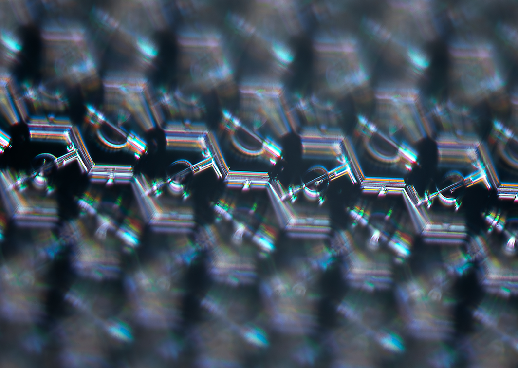
Closeup of the Science Eye, a visual prosthetic device to restore sightCloseup of the Science Eye, a visual prosthetic device to restore sight

Science Eye

Offering new hope for patients with serious blindness due to photoreceptor loss

In some diseases like retinitis pigmentosa and macular degeneration, the light-sensitive cells of the retina, the photoreceptors, are lost while the cells of the optic nerve, the retinal ganglion cells, remain.

Since the ganglion cells are the output of the retina, if we can stimulate them directly, we can restore visual input to the brain even without replacing the photoreceptors.

Closeup of the Science Eye, a visual prosthetic device to restore sight

The retina converts light into neural signals for processing by the brain

Photoreceptors are light-sensitive cells that absorb light and convert it into information

Once light is converted into information, it gets passed onto a layer of intermediate neurons

The intermediate neurons connect to the retinal ganglion cells which project into the brain and form the optic nerve

A medical diagram magnifying the different layers of the retina
The Science Eye is a "combination device" with two key components:
Optogenetic gene therapy

A protein nanoparticle is used to deliver a gene to some of the retinal ganglion (optic nerve) cells, making them light-sensitive at a specific wavelength.

A colorized render of a spiky-looking protein nanoparticle
High-resolution display film

A tiny, flexible high-resolution display film is surgically inserted over the retina to allow fine control of the newly light-sensitive ganglion cells.

A diagram of an LED array curving to fit against the RGC layer of the retina

Size comparison

The electronics package is similar in size to glaucoma shunts widely used today, which are routinely inserted without general anesthesia and can't be felt by patients once inserted.

  • Science Electronic PackageA render of the electronics package of the Science Eye on the outside of an eyeball, for size comparison
  • Ahmed ValveA render of an Ahmed glaucoma valve on the outside of an eyeball, for size comparison
  • Baerveldt ShuntA render of a Baerveldt glaucoma shunt on the outside of an eyeball, for size comparison
Side swipe to see more

Glasses

Glasses worn by the user contain sensors, processing, and batteries which supply power and data to the implant.

  1. Resonant inductive coil for wireless power

  2. Binocular cameras

  3. Infrared optical data connection with implant

Receiving a Science Eye is a straightforward three-part procedure
A render of the Science Eye unfolded against the outside of an eyeball

The implant is slid in over the eye and the display is inserted through a small incision

A render of the Science Eye inserted into an eyeball

The implant is secured in place and the display is laid on the retina

A render of the Science Eye inserted and secured against the inside of an eyeball

The display film is affixed to the retina to hold it in place

Stay up to date with our progress

I am a:

Condition